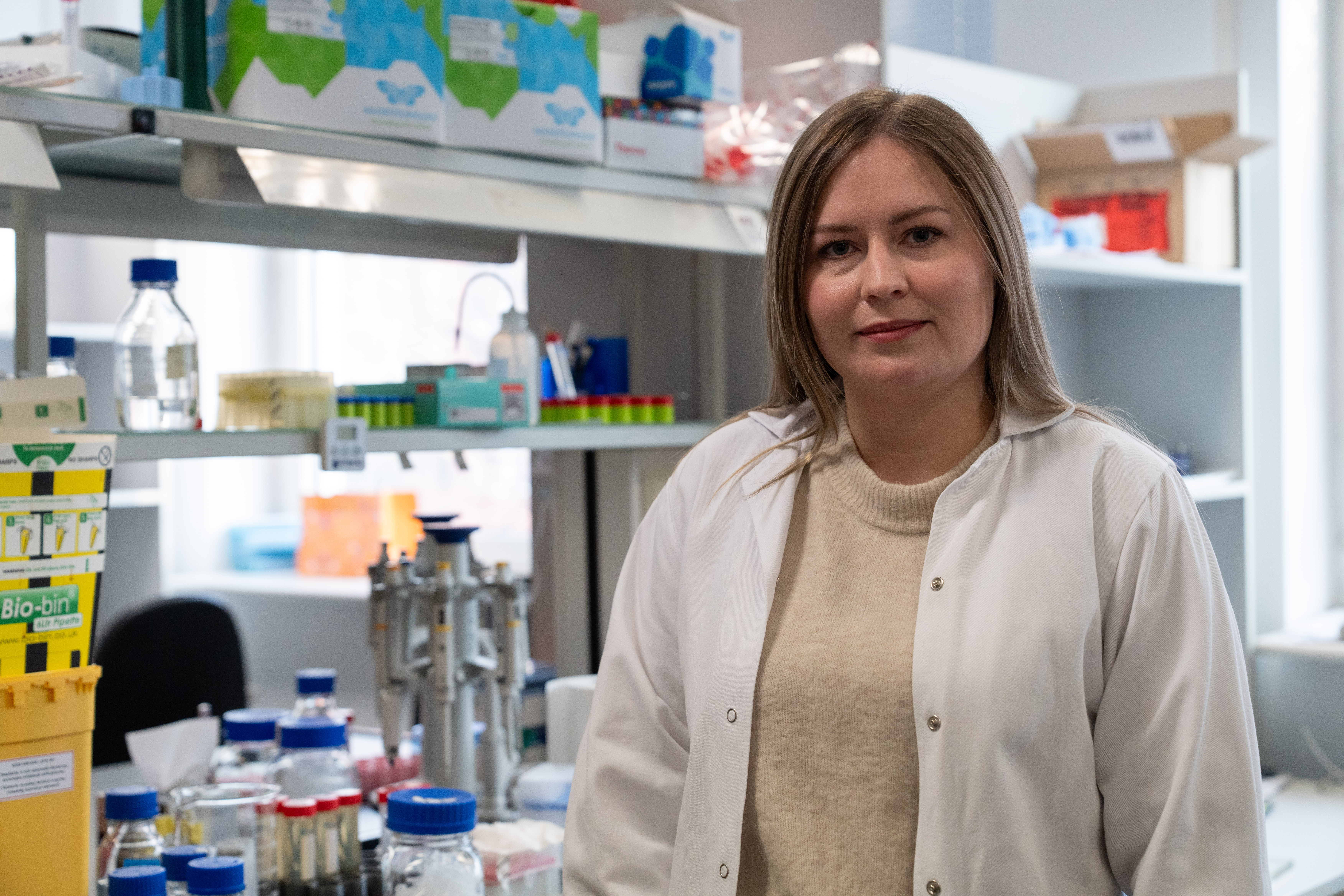
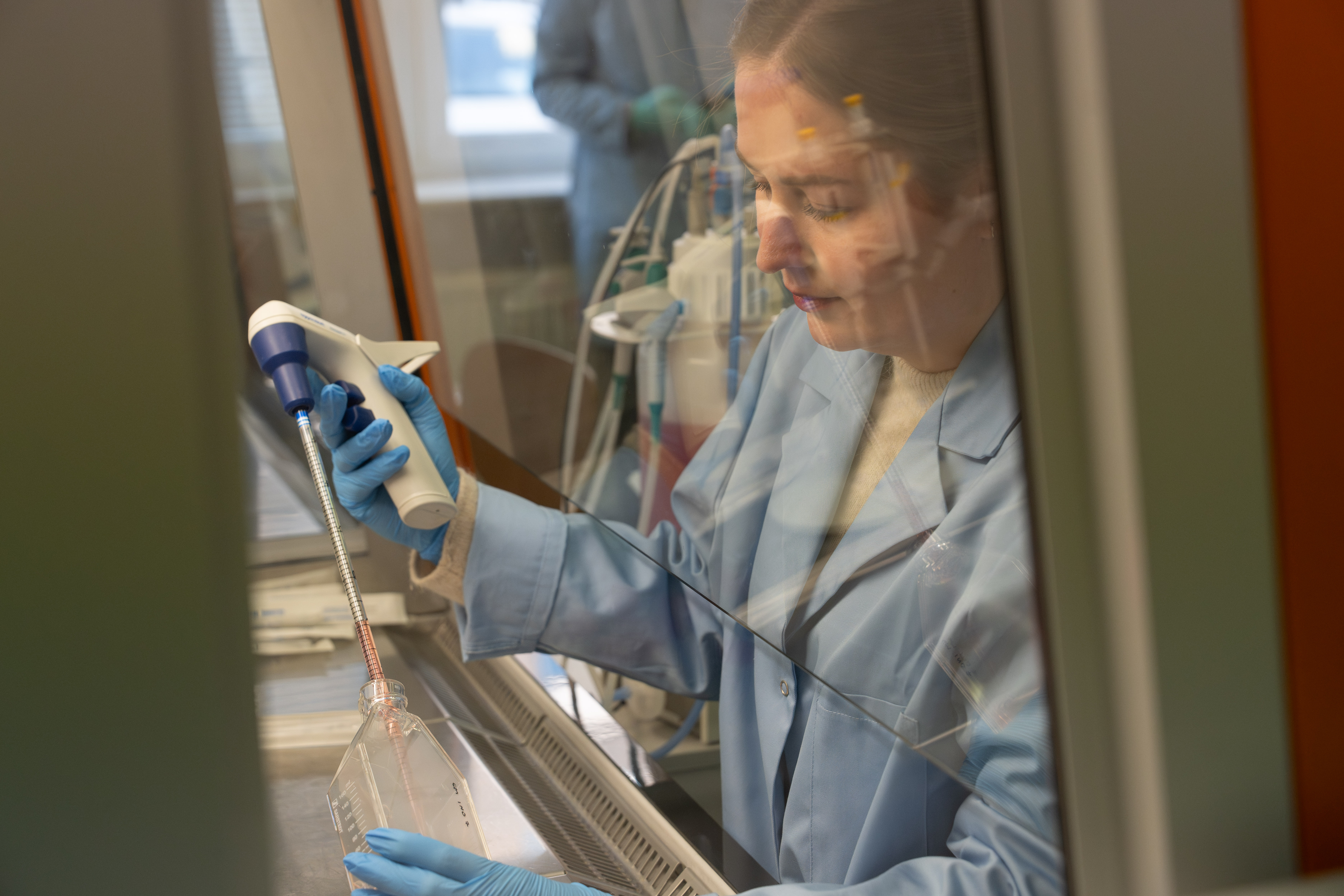
Breast cancer is the most common cancer in women. It is estimated that nearly 1.7 million patients are diagnosed with it each year, and over 500,000 die from it. In Poland, breast cancer has become one of the greatest threats causing premature mortality in women over the last century. Dr Justyna Topa from the Intercollegiate Faculty of Biotechnology UG and MUG has contributed to research on this cancer by focusing her doctoral thesis on circulating tumour cells (which cause metastases) with different phenotypes in breast cancer patients. For her doctoral thesis, she received the Rector's Special Award ‘I include gender in research’.
Urszula Abucewicz: - Every day, as many as 55 Polish women hear the diagnosis: breast cancer. Every year, more than 20,000 women are diagnosed with it, and about 9,000 die.
Dr Justyna Topa: - Breast cancer is a very serious and extremely common global problem. Statistics show that one in eight women will develop breast cancer during their lifetime. Fortunately, the prognosis for patients diagnosed early is very promising. The chances of survival decrease when distant metastases occur.
- How do metastases develop?
- Metastases originate from cancer cells that have separated from the primary tumour in the breast, entered the bloodstream and, after finding a favourable niche, were able to adapt, divide and lead to the formation of new cancerous lesions. The cells that cause metastases were the subject of my research during my doctoral thesis. We call them circulating tumour cells (CTCs). They are very clever; they can deceive the immune system by effectively masking themselves or sending signals that identify them as ‘non-threatening’.
- Why did you focus on them?
- Cancer cells circulating in the blood can be a biomarker in the early stages of the disease. The mere presence of these cells in the blood of patients gives us information about the possible progression of the disease. In my doctoral thesis, I focused primarily on their characteristics at the mRNA and protein level.
- What did this research involve?
- In a prospective study, I analysed samples from 104 patients with early-stage breast cancer, with small tumours, no metastases, but preferably with affected lymph nodes.
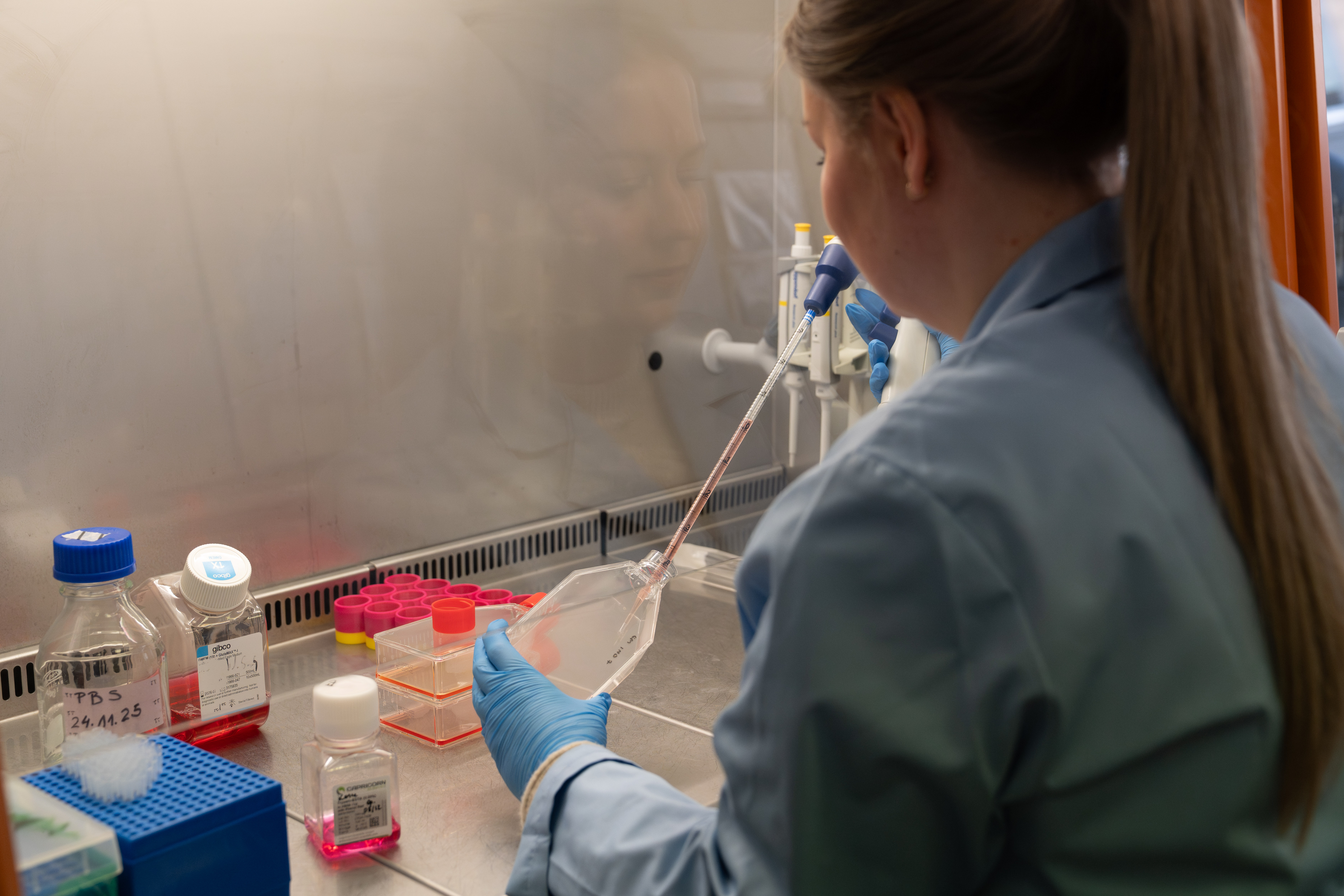
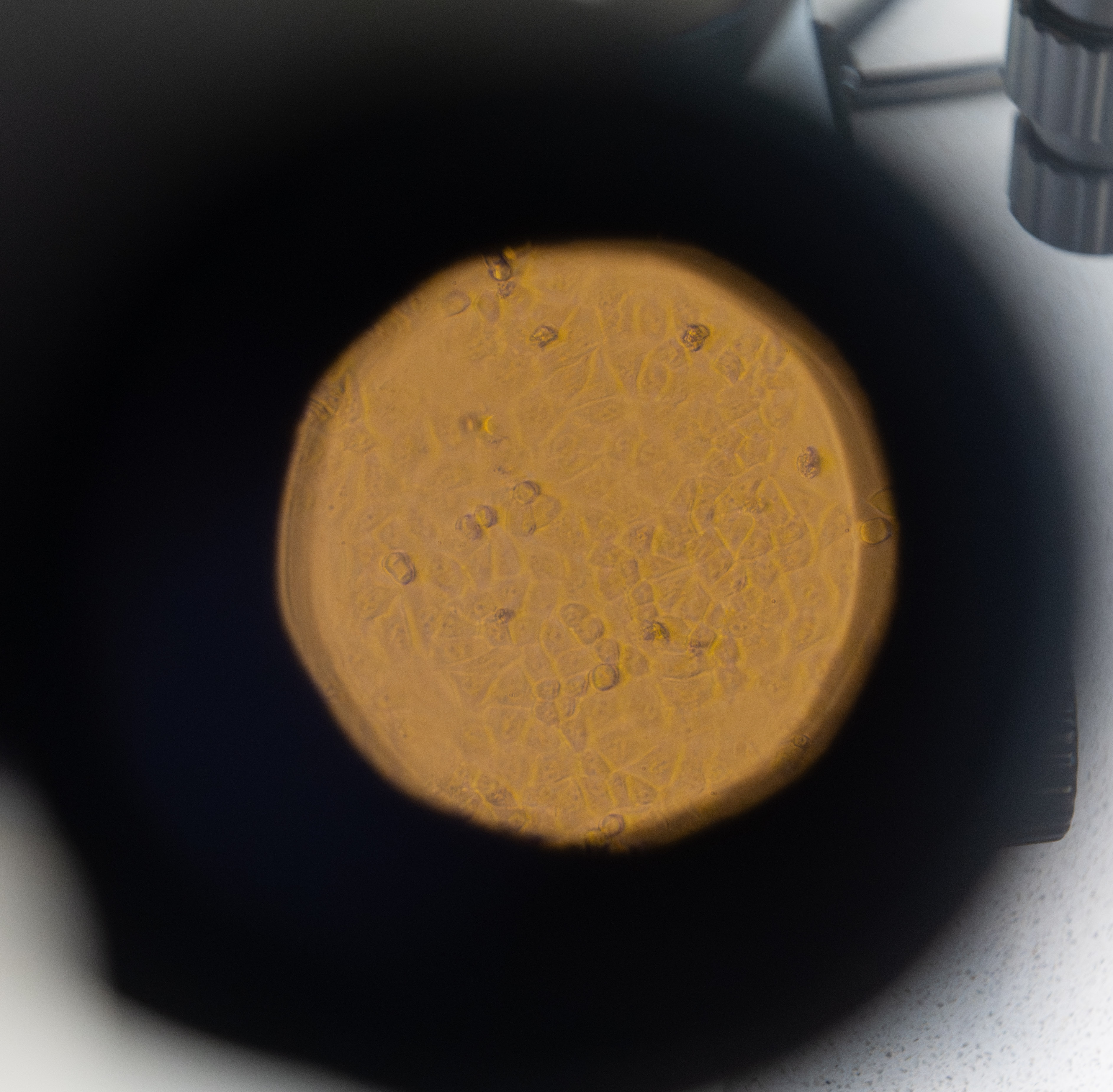
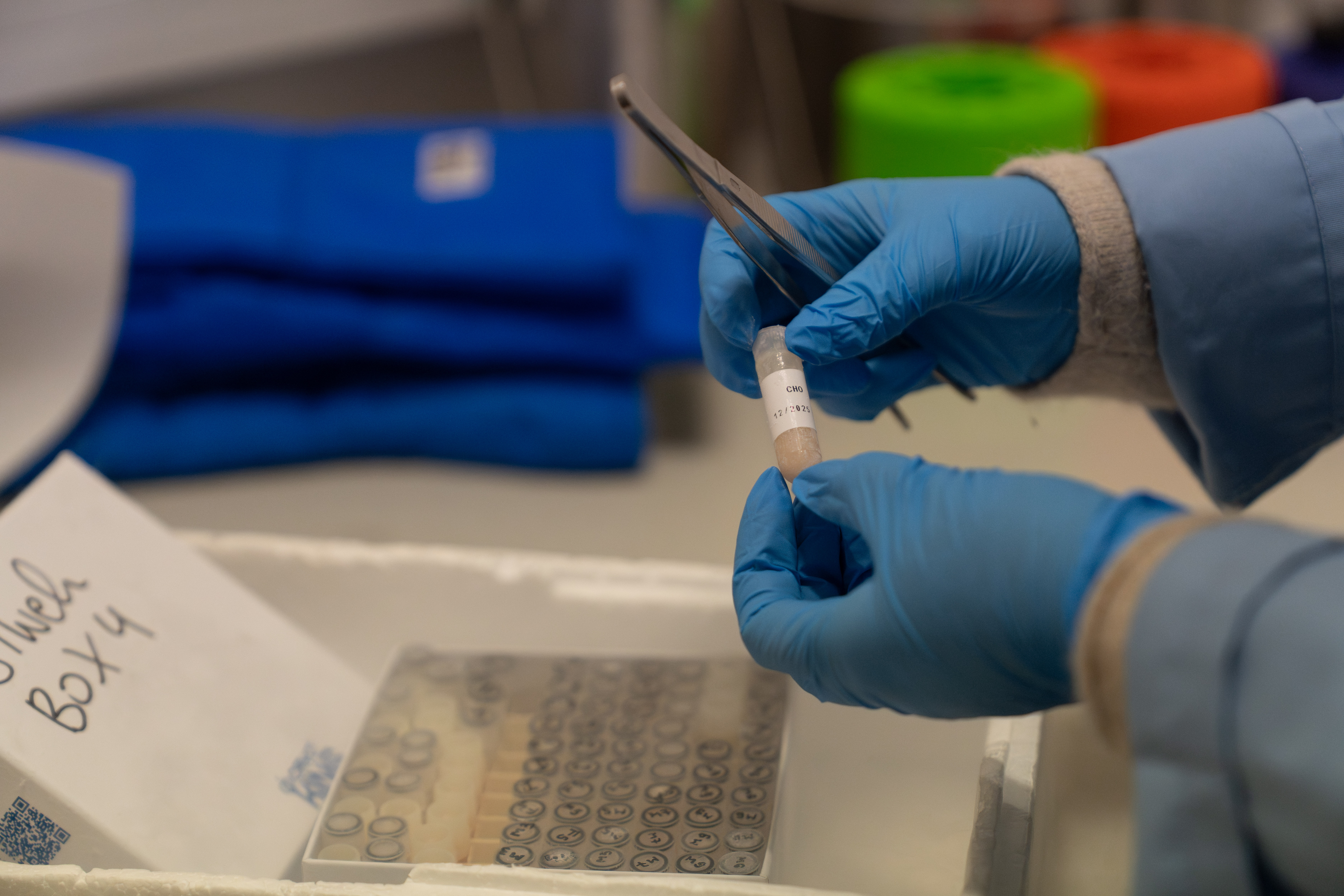
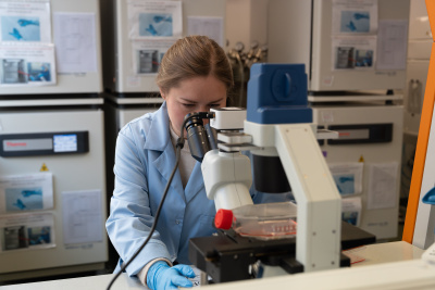
My task was to capture CTCs, which are very rare in the early stages of the disease. The challenge was to sift through billions of immune system cells and manually isolate individual CTCs, which I then characterised. In a single blood sample from a patient, I found only a few, usually just one cell of interest. The bloodstream is not a natural environment for CTCs. Most of them survive only a few hours, and only a small percentage can initiate metastasis in the future.
- So you were looking for a needle in a haystack.
- You could say that.
- The ones that have undergone epithelial-mesenchymal transition, or, to put it simply, have changed their lifestyle and become mobile instead of static, are more dangerous or more aggressive, right? Their migration through the body can lead to metastasis.
- In breast cancer, all cancer cells are initially epithelial cells. Epithelial cells naturally form compact clusters and remain in close contact with other cells and the substrate. As a result of epithelial-mesenchymal transition (EMT), cancer cells can lose their epithelial characteristics and acquire mesenchymal properties. This is accompanied by a change in cell shape, loss of the need for constant contact with other cells and the substrate, and increased plasticity.
As a result of EMT, cancer cells become more independent. They can separate from a group of other cancer cells, make their way through the tumour microenvironment and cross the blood vessel barrier. In this way, cancer cells enter the bloodstream, from where they can travel to distant tissues and organs, where they can initiate the process of metastasis.
Until now, cancer cells circulating in the bloodstream have been a blank slate for researchers. We knew little about what happens to them, how they survive and reach another organ to form metastases. However, we do know that CTCs that have already undergone EMT, upon reaching the site of metastasis, reverse this process and become more epithelial, i.e. more epithelial, and thus can more easily settle in new locations.
This does not mean, however, that only cells after EMT can metastasise. During my research, I wanted to find out what stage of EMT CTCs isolated from the bloodstream are in, and I was particularly interested in cells with an intermediate phenotype - epithelial-mesenchymal. According to the literature and previous research by the team, it is these cells that often exhibit characteristics conducive to survival and aggressive behaviour.
- Is the presence of circulating tumour cells in the blood tested during the diagnosis and treatment of breast cancer?
- The US Food and Drug Administration (FDA) has approved two methods for detecting circulating tumour cells, but they are not currently used for routine diagnosis of breast cancer patients. The first, CellSearch, is a method that detects circulating tumour cells based on the presence of epithelial markers - EpCAM and cytokeratins - and the absence of the blood cell marker CD45. However, this method overlooks cells that have lost the expression of epithelial markers and gained mesenchymal characteristics. It is suspected that it is precisely cells with this phenotype that are more resistant to stress, more easily evade immune system control and, above all, have characteristics that help them leave the primary tumour.
The second method, approved by the FDA nearly three years ago, utilises the difference in size between cancer cells and immune system cells in the blood. However, it should be noted that we still need a suitable marker to identify these CTCs. We have very well-defined epithelial markers, but we still lack a marker for cancer cells that have undergone complete EMT. What I mean here is the lack of a marker on the cell surface that would allow us to easily identify these cells while they are still alive. This is important because the analysis of living cells allows us to learn about their transcriptome (mRNA), which tells us which genes are being expressed.
- Let's go back to the first method. It's a bit like looking for people in suits who have taken off their jackets.
- Each cell has its own suit of characteristic features. We call it a phenotype. In the case of breast cancer cells, we distinguish between epithelial and mesenchymal phenotypes, but this is not a binary division.
In my research, I also identified an intermediate phenotype - epithelial-mesenchymal. It turns out that cells with this phenotype, despite retaining their epitheliality, exhibit characteristics indicative of their aggressiveness, which is characteristic of cells with a mesenchymal phenotype. The heterogeneity of cancer cells with different phenotypes and properties is actually the biggest challenge, as we are looking for very different cells with unique characteristics that could have changed them when they left the primary tumour and entered the bloodstream.
The CellSearch method allows only those cells that have retained their epithelial ‘outfit’, i.e. characteristics typical of epithelial cells, to be detected. In practice, this means that only some CTCs are detected. However, it should be remembered that during their ‘journey’, cancer cells can change their phenotype, which also results in a change in the way they function. This change does not always result from the formation of new mutations. Very often, it is the result of adaptation to a changing environment. The cell does not need to modify its genome - it is enough for it to change the set of proteins it produces, adapting to the current conditions.
- Circulating tumour cells can get dormant and hide.
- The patient feels well, the treatment seems to be effective, and doctors assume that the disease has been controlled, while individual cancer cells may enter a state of dormancy. Such cells can remain in a given organ or tissue for a long time, without dividing or showing any symptoms. They wait for favourable conditions to reactivate, begin to divide, and, as a result, lead to metastasis. Individual cancer cells in a metastatic niche are invisible to standard diagnostic methods with limited sensitivity. Such foci are only detected when the mass of cells reaches a certain size. This is why it is so important to understand what happens between the cancer cells leaving the primary tumour and the appearance of metastases. In my research, I wanted to focus on single circulating tumour cells because they are what cause the spread of cancer. I wanted to understand what makes them unique: what characteristics allow them to leave the tumour, survive in a very hostile environment and potentially initiate a new cancerous lesion elsewhere.
Perhaps in the future, based on these characteristic features, it will be possible to design new therapies - although it must be emphasised that this is a very distant prospect. One of the biggest obstacles is the fact that circulating tumour cells are extremely rare and, at the same time, very biologically ‘experienced’. In the bloodstream, they are exposed to enormous stress and, in addition, they must survive the isolation process itself, which is very stressful for them. As a result, these cells are often in poor biological condition, which makes further research on them very difficult. Therefore, the first and crucial step is to develop the best possible methods for isolating them - methods that are fast, effective and as non-invasive as possible.
Only in the next stage is it possible to profile them accurately and attempt to identify characteristic markers, i.e. features that would allow them to be detected - whether in the blood or perhaps in tissues.
The profiling of circulating tumour cells alone can already help in discovering new potential therapeutic targets. In my research, despite the very limited number of cells meeting the quality criteria, I observed that in cancer cells with mesenchymal characteristics, the process of protein formation is limited. This is a mechanism that provides them with the absolute minimum necessary for survival and gives them a chance for further expansion in the future. Perhaps it is precisely the disruption of pro-survival pathways that could become the basis for targeted therapy in the future. This is crucial for the treatment to be as precise as possible and, at the same time, as least burdensome as possible for the entire body.
- How do oncologists view the results of your research? How do they react to the information that circulating tumour cells (CTCs) are detected in a significant proportion of patients?
- My research covered a relatively small group of patients in the early stages of the disease - just over a hundred. In order to draw clear clinical conclusions, much larger studies are needed, preferably multicentre studies involving hundreds or thousands of patients. The first results of a large clinical study, STIC CTC, which involved several hundred patients, are already known. It examined whether the selection of therapy based on the number of circulating tumour cells in the blood can compete with the currently used criteria for assessing patients. Studies published in recent years indicate that determining the number of CTCs in the blood provides prognostic information comparable to the diagnostic criteria used to date.
In this context, my research is rather a small ‘brick’ added to a much larger puzzle. Its value lies, among other things, in the fact that both my results and those of other research teams are publicly available. This allows them to be combined, analysed on larger data sets and checked to see what conclusions emerge when the number of cells or patients analysed is increased. At this stage, this is my role - to provide data that can be used in larger projects in the future. It should be emphasised that this is basic research, not clinical research. However, it is on the basis of such research that the design of large clinical trials begins, which can have a real impact on medical practice.
When it comes to interpreting the results, I would not approach them in a black-and-white manner. All cancer cells - regardless of phenotype - that enter the bloodstream pose a potential threat. In analyses based solely on the presence of CTCs, we have indeed observed that their detection correlates with worse clinical and pathological features of the tumour. Only single-cell analysis revealed something more: cells that have acquired mesenchymal characteristics - both purely mesenchymal and with an intermediate epithelial-mesenchymal phenotype - enter a pro-survival state. They are, in a sense, ‘geared’ towards survival in difficult conditions. They also have their limitations. Cells with strong mesenchymal characteristics find it more difficult to initiate metastasis because, in order to do so, they must switch from a pro-survival mode to a mode of active proliferation, i.e. a mode of intensive multiplication. Simply escaping from the tumour and surviving in the bloodstream is not enough. The key is whether the cell can ‘complete the process’, i.e. start forming a new tumour. For this to happen, it must adapt to a completely new environment. Numerous challenges await it in the new tissue: the presence of immune system cells, different metabolic conditions and environmental signals than in the primary tumour. Only a small proportion of cells are able to adapt to this and give rise to distant metastasis.
- Are you continuing your research on CTCs?
- Dr hab. Aleksandra Markiewicz, my doctoral supervisor, is conducting further intensive research in this area. She is continuing her research on circulating tumour cells, expanding it not only to include transcriptome analysis, which I was involved in, but also genome analysis. At the same time, further grant applications are being submitted and the research is being systematically developed.
- What are you working on now?
- I am currently researching the phenomenon of transcriptional memory. This is a process in which a cell treated with a specific factor ‘remembers’ this signal and, upon subsequent contact with it, responds with significantly stronger gene expression at the mRNA and protein level. My task is to investigate what causes this memory and what its mechanisms are.
At the end of last year, I was awarded a grant in the NSC MINIATURA competition, under which I am investigating the cumulative effect of transcriptional memory depending on the number of exposures to a given stimulus. In my research, this stimulus is interferon gamma, a protein that plays a key role in the body's immune response. I am analysing how repeated stimulation with interferon affects cell behaviour.
The research is already at an advanced stage, and in the future, I would like to try to apply its results in cancer therapy. I also have an idea to combine this research with the topic of my doctoral thesis, i.e. circulating tumour cells. This is still a concept that needs further refinement. It has only been a year since I defended my doctoral thesis, and I feel that I still need time to learn, observe and gain scientific maturity. However, I hope that in the future, the results of this work will not only be published but will also be valuable for new solutions in clinical practice.
The doctoral dissertation of dr Justyna Topa from the Intercollegiate Faculty of Biotechnology UG and MUG, entitled ‘The clinical and biological significance of circulating tumour cells with different epithelial-mesenchymal phenotypes in breast cancer patients’, was prepared under the supervision of prof. dr hab. Anna Żaczek and dr hab. Aleksandra Markiewicz.